
Discover HOLOEYE Photonics AG
HOLOEYE is a leading provider for design, development and commercialization of industrial micro display applications, adaptive micro-optics, Spatial Light Modulators and diffractive components. With our specialized, flexible technologies, our tailor-made solutions and our extensive know-how, we set new standards and enable groundbreaking applications in R&D and industrial integration.
Customized Product Developments
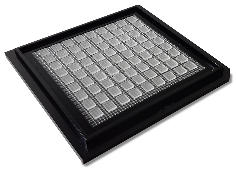
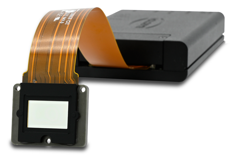
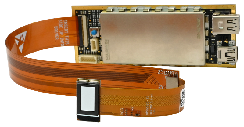
HOLOEYE offers comprehensive customization and development services, no matter if based on phase modulation Spatial Light Modulators, LCOS microdisplay components for monochrome or color projection applications, standard Diffractive Optical Elements or developments of a Diffractive Optics solutions from scatch.


phase

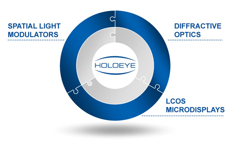
About HOLOEYE
HOLOEYE is providing products in the fields of Spatial Light Modulators, Diffractive Optical Elements and LCOS microdisplay components.
We provide highly specialized, most flexible and diversified standard solutions for academic and industrial R&D and offer the highest level of component customization, custom developments and volume production for industrial integration.
News
Stay up-to-date with the latest events and exciting developments at our company. In this dedicated news section, we’ll keep you informed about our innovative products, industry partnerships, noteworthy achievements, and much more.
Current Events
Get information about the upcoming industry exhibitions, trade shows, conferences, webinars, and other events where HOLOEYE will be showcasing our latest innovations and solutions. Meet us face-to-face and interact with our knowledgeable team, experience our products firsthand, and gain valuable insights into the cutting-edge technologies we offer.
We look forward to meeting you at our next event!